Struvite Bladder Stones in Singapore dogs
Dr Sing KongYuen, BVMS (Glasgow), MRCVS
Toa Payoh Vets
July 31, 2012
Difficulty in peeing, not able to pee and peeing urine with blood are the most common reasons for dog owners to seek veterinary advices at Toa Payoh Vets. Sometimes, the owner sees stones passed out in the urine.
Urolithiasis in dogs is such a large topic of a few hundred pages as there are several types of bladder stones affecting the dog and their diagnosis and treatment vary. Therefore, only struvite urinary stones, being most commonly seen at Toa Payoh Vets will be discussed in this article.
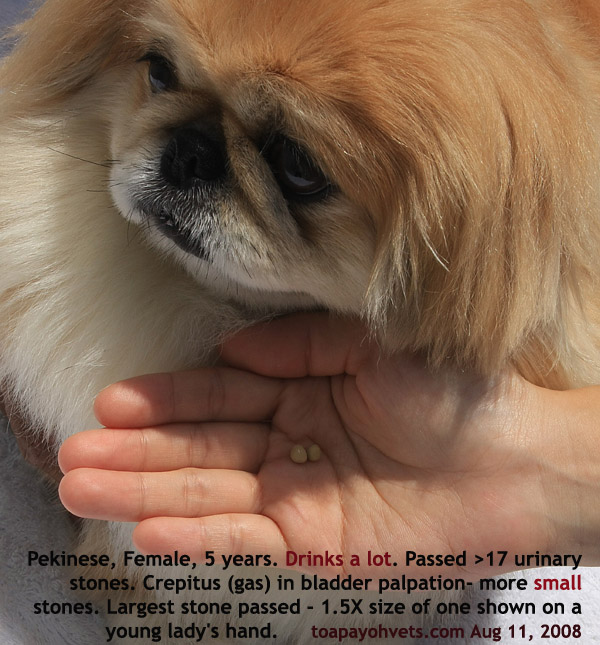
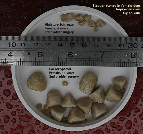
Breeds usually affected include the Miniature Schnauzer, Shih Tzu, Bichon Frise, Miniature Poodle, Cocker Spaniel and Lhasa Apso, but any breed can be affected. Female dogs are said to form approximately 85% of bladder stone cases.
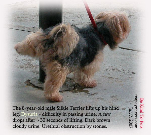
Some dogs may not show clinical signs of blood in the urine, difficulty in urination or inability to urinate until much later in the disease with severity of signs depending on the location, size,and number of uroliths formed. These stones can be formed anywhere along the urinary tract in the kidneys, the urethra and the bladder.
Struvite stones are composed primarily of Magnesium, Ammonium and Phosphate (MAP). They are formed within the urinary tract and occur when the urine is supersaturated with MAP (i.e. large quantities of the crystals are present). MAP supersaturation of urine may be associated with several factors, including urinary tract infections, alkalineurine, genetic predisposition and diet.
Your vet will take a comprehensive history to determine the commencement and the severity of the disease. Physical examination include bladder palpation to feel the crepitus (sounds of gas and stones rubbing against each other) inside the bladder or the solid stones if they are large.
However, a complete blockage of the urinary tract is life-threatening as the dog can't pee and the full bladder may rupture with delays in treatment. In such cases, a urinary catheter will be used to unblock the obstruction or the urine is extracted via the bladder as soon as possible. This is done to protect the bladder and kidney from further damage.
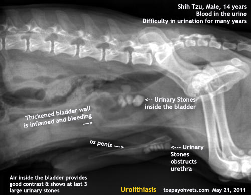 X-ray of a dog that cannot pee (left). If the dog cannot pee, the likely cause is urethral obstruction caused by urinary stones being stuck inside the urethra.
Blood screening, urine analysis and radiographs are usually performed to confirm the presence of urinary stones. Abnormal blood work may show if the obstruction of the urinary tract is severe. Blood tests may show changes to the kidney function or an increase in white blood cell counts affecting the health of the dog.
Urine analysis is the most useful and should always be done. A sterile sample is taken either via catherisation (passing a tube into the bladder) or cystocentesis (straight from the bladder). With the urine sample analysed, MAP crystals can be present but this is not always the case. For example, stones that are too well formed or too large may not shed crystals. Therefore, the vet should not deem the absence of crystals in the urine as no struvite or urinary stones being present in the affected dog.
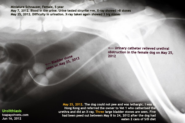 X-ray of a catheter to push back the stones into the bladder in a female dog that could not pee at all as the stone was stuck inside the urethra
In addition, urine pH gives the vet a good idea of the nature of the stone. Struvite crystals are formed very commonly in an alkaline environment in which bacteria is present. A urine sample can show the presence of bacteria. The bacteria be cultured to know the type of bacteria causing the infection and antibiotic sensitivity tests can be performed by the laboratory to advise on the appropriate antibiotics to be prescribed.
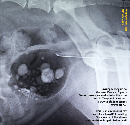 Struvite uroliths are radio-dense and can be detected on radiographs.
 However, they need to be of a certain size before they are evident. The number and size of urinary stones seen in the X-ray may not correlate with the severity of clinical signs.
However, a radiograph is highly recommended for the vet to know the number and size of stones and where they are located prior to surgical removal, if surgery is to be advised.
Clients need to understand that in spite of all the tests above, the composition of the actual stone cannot be determined unless a stone sample (from the surgery or that has been urinated out) is sent to the laboratory for analysis.
Treatment options for struvite bladder stones revolve around surgery or medical dissolution of the stones.
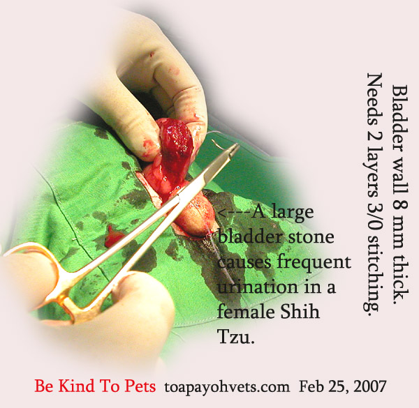
Benefits of surgery include faster recovery times, and the ability to identify the actual type of stone involved. Surgery is indicated if the stones are too large or too well formed as they may not dissolve medically. Disadvantages of surgery are that it is more invasive and there are risks associated with general anesthesia in a sick and/or older dog.
For clients that are not so comfortable with surgery or in cases where surgery is not advised due to health concerns (e.g. the dog is very old and in poor health), the alternative is medical dissolution. This medical solution is non-invasive but takes a much longer time to show the effect as the stones are dissolved slowly. However, large stones may not dissolve at all. One important note to take into consideration is that there is no way to accurately determine the nature of the stone without sending it for laboratory analysis. Obviously, the medical solution is not applicable to all types of urinary stones (e.g. calcium oxalate stones), but it is especially effective and useful in struvite stone dissolution.
Medical dissolution revolves around 3 main concepts. They are to acidify the urine, to reduce the intake of MAP such that it does not saturate in the urine and to dilute the urine so crystals do not have a chance to form. For struvite uroliths, there are specially formulated diets such as the Canine S/D, C/D or W/D that I have used to dissolve the stones.
Medical dissolution of stones takes a mean time of 3 months. The time taken for complete dissolution is varied depending onthe size of the uroliths and the quantity. Severe cases can take up to 6 months before the stones are fully dissolved. However, very large stones will not dissolve.
Along with this diet change, I prescribe an appropriate antibiotic course to treat any primary or secondary bacterial infection. During treatment, only the prescription diet should be used. I usually advise no dog treats or other food and to encourage the dog to drink water.
The S/D diet is used initially for 1-6 months before switching over to the C/D or W/D diet. It is not recommended for:
1. Use concurrently with urinary acidifiers
2. Feeding longer than 6 months
3. Dogs with non-struvite uroliths (urinary stones).
Transition to feeding S/D should be done over a period of seven days, gradually introducing the amounts during the transition period and monitoring the patient. Most dogs will not eat the S/D diet immediately and so the owner must be educated to switch to the S/D diet gradually over at least 7 days.
After successful dissolution of struvite stones confirmed by urine analysis and X-rays, Canine C/D or W/D can be used for maintenance. Canine S/D should not be used for the prevention of bladder stones as the diet is low in MAP and protein. Long term use of this diet is not recommended as the nutrients are not be sufficient.
Key benefits of Canine S/D include:
· Low levels of MAP to aid in dissolution of struvite uroliths and crystals.
· Promotion of acid urine by reducing the urinary pH to 5.9-6.1 (targeted) to increase the solubility of struvite crystals.
· Lower protein levels result in increased urine volume and more dilute urine.
· Antioxidants are added to defend cells from free radicals and to promote a healthy immune system.
In this article, I have written about the S/D, C/D and W/D prescription diets for the medical treatment of struvite urinary stones as I have used them in my practice. However, there are other equivalent prescription diets from other manufacturers and it is up to your vet to advise you as to the type of prescription diet to use or to get bladder surgery done to resolve the problem fast.
Many Singapore dog owners do not adopt my advices to review the cases 1-3 monthly and do urine tests and X-rays to ensure that no new stones are formed after surgical removal of the stones or after using the S/D diet. They are happy to see that the dog has not passed blood in the urine and does not have difficulty in urination and continue with feeding the usual dry dog food again.
In some cases, the problem recurs and it can be heart-breaking and costly if another surgery is required. So some owners elect to euthanase the dog. Regular urine tests would have been most useful in detecting the presence of struvite stones although the absence of struvite crystals in the urine does not mean that there are no stones present. Only X-rays will be able to tell. Sometimes, a dog that has had struvite stones may become later affected with another type of stone such as calcium oxalate stones and that is why regular urine tests are so important.

In conclusion, be alert as to the urination pattern of your older dog and seek veterinary advice promptly if there are signs of discoloured urine, urinary difficulty or inability to pee.
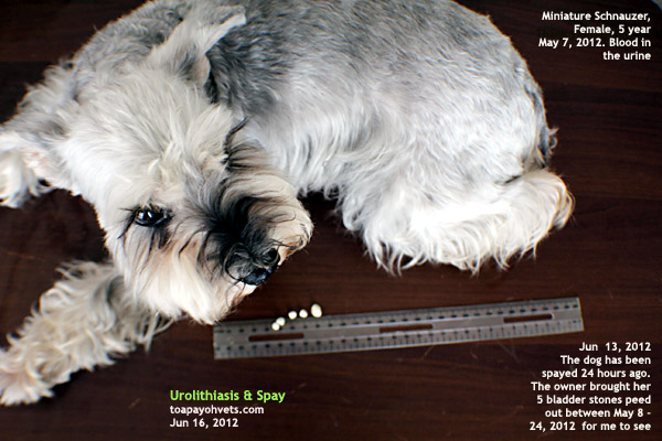
Image of a female dog that has peed out urinary stones
For more detailed case studies of urinary stone cases seen at Toa Payoh Vets, goto:
http://www.bekindtopets.com/animals/20081201PG7_Dog_Surgery_Anaesthesia_Urinary_Tract_Problems_ToaPayohVets.htm
Acknowledgement: I thank Dr Daniel Sing for his contribution to this article and various dog owners for permitting me to record their cases in this article