Article
http://www.medirabbit.com/EN/Eye_diseases/Disorder/Lipid/Lipid_en.htm
Corneal lipidosis - also called corneal dystrophy or lipid keratopathy - is a condition where excess lipids (usually cholesterol esters) or minerals (calcium) are deposited under the surface of the cornea. The infiltration usually starts at the edge of the cornea and can be observed in the anterior stroma, the epithelial basement membrane and the epithelium.
Corneal lipidosis is not associated to a disease; it is not breed or gender dependent.
Etiology
A lipid rich diet and/or trauma are the main causes for lipid deposits into the cornea. Congenital factors cannot be ruled out.
Clinical signs and diagnosis
It is based on a complete ophthalmic examination and a discussion with the owner about the food fed to the rabbit.
Both eyes are usually affected (bilateral) but not necessarily to the same degree. Unilateral lipidosis has rarely been reported. The fat deposits, which usually start near the third eyelid, can be opaque, raised, subtle and pale, bright white, silver or grey colored areas. Vascularization is observed in the affected part of the cornea. While the cornea is mainly affected, fat deposits have also been noted in the lens, iris and ciliary body of a Dutch rabbit. Often it is accompanied by macrophage invasion. An inflammatory process has been observed, but does not always seem to be present.
Unlike in dogs, corneal lipidosis is associated to gradual loss of vision in rabbits. If the deposit is severe, it can lead to ulceration of the cornea.
There is no pain associated to this condition.
http://circ.ahajournals.org/cgi/reprint/18/4/519Lipid plaque in hypercholesteremic rabbit cornea occurring at site of vascularity. Cornea had been cauterized several times by heated probe. | http://circ.ahajournals.org/cgi/reprint/18/4/519Sections of rabbit cornea in region of plaque stained with hematoxylin-Sudan. Noteworthy is abundance of intracellular globular lipid and relatively slight amount of granular sudanophilia. |


Treatment
There is no medical therapy available, other than bring modification to the diet. Fatty food and milk-based products (cheese, butter, and yogurt) should be discontinued.
It is not known if superficial keratotomy can help.
Further information
Fallon MT, Reinhard MK, DaRif CA, Schoeb TR. Diagnostic exercise: eye lesions in a rabbit. Lab Anim Sci. 1988; 38:612-3.
Garibaldi BA, Goad ME. Lipid keratopathy in the Watanabe (WHHL) rabbit. Vet Pathol. 1988; 25:173-4.
Gwin RM, Gelatt KN. Bilateral ocular lipidosis in a cottontail rabbit fed an all-milk diet. J Am Vet Med Assoc. 1977; 171:887-9.
Hillyer E.V., Quesenberry K.E., Ferrets, Rabbits, and Rodents: Clinical Medicine and Surgery, New York: WB Saunders Co., 1997, p. 339-345.
Sebesteny A, Sheraidah GA, Trevan DJ, Alexander RA, Ahmed AI. Lipid keratopathy and atheromatosis in an SPF laboratory rabbit colony attributable to diet. Lab Anim. 1985; 19:180-8.
Stock EL, Mendelsohn AD, Lo GG, Ghosh S, O'Grady RB. Lipid keratopathy in rabbits. An animal model system. Arch Ophthalmol. 1985 May;103(5):726-30.
---------------------------
Procedure for corneal ulceration examination
1. Schirmer tear test
2. Assess corneal and palpebral reflex
3. Thorough examination of the eyelid and conjunctival anatomy and function including
the caudal surface of the 3rd eyelid.
4. Microbiological examination if corneal ulcer is believed to be infected.
5. Fluorescein staining
------------------------------------------------------------
video rabbit with corneal ulcer fluorescein stain test


-----------------------------------------------------------------------------
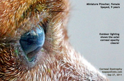
DOG CORNEAL DYSTROPHY


-------------------------
Normal human eye cornea

No comments:
Post a Comment
Note: Only a member of this blog may post a comment.